
Research-grade optics
High-resolution infinity optics and suitable high-NA objectives support detailed observation of cells, tissue sections, chromosomes and fluorescent biological targets.
Biological fluorescence microscope
A high-end modular research fluorescence microscope for demanding life-science imaging, including FISH, karyotyping, cytogenetics, immunofluorescence, cell biology, pathology and molecular biology workflows.

Designed for research
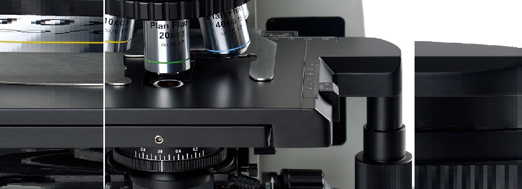
The BM-R’s large, double-way linear-drive stage can be controlled from either side and supports two slides for comparative study. Its anti-corrosive, anti-friction surface and arc-transition platform help improve durability and protect against impact.
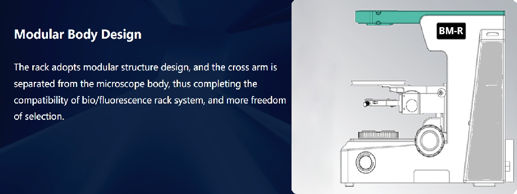
A modular frame separates the cross arm from the main body, improving compatibility between biological and fluorescence systems. Low-position coaxial controls, adjustable tension and an upper limit stop provide accurate focusing and comfortable extended use.
Multiple trinocular light-splitting options direct light to the eyepieces, camera, or both simultaneously. Inverted- and erect-image heads are available for observation, documentation and motorized workflows.
Advanced fluorescence imaging
The BM-R can combine infinity-corrected research optics, high-numerical-aperture objectives, efficient epi-fluorescence illumination, precision mechanics and sensitive digital imaging. Each system is configured around the fluorophores, specimens, observation methods and automation requirements of the laboratory.
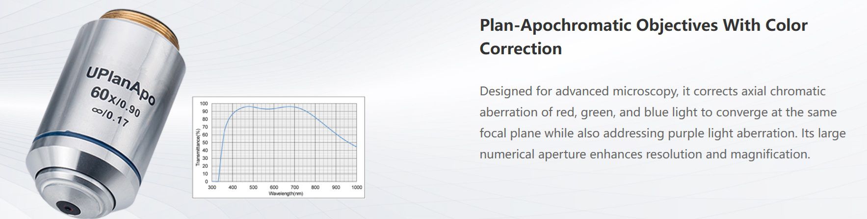
High-resolution infinity optics and suitable high-NA objectives support detailed observation of cells, tissue sections, chromosomes and fluorescent biological targets.
Filter configurations can support UV, blue, green, yellow, orange, red and far-red fluorescence according to the laboratory's fluorophores and application.
Choose a monochrome scientific camera for demanding fluorescence detection or a colour camera for brightfield, histology and routine biological documentation.
Manual or motorized focusing supports accurate work with thin specimens and can enable repeatable Z-stack acquisition with compatible imaging software.
Optional motorized nosepiece, focus, XY stage, fluorescence selection and illumination control support multichannel, multi-position and automated imaging.
Add fluorescence channels, objectives, scientific cameras, motorized components and analysis software as research requirements develop.
FISH and chromosome imaging
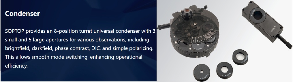
Fluorescence In Situ Hybridization uses fluorescently labelled probes to identify DNA sequences within chromosomes or cells. A suitable BM-R configuration combines stable focusing, high-resolution optics, efficient illumination, accurate channel separation and a sensitive camera.
Fluorescence optical pathway
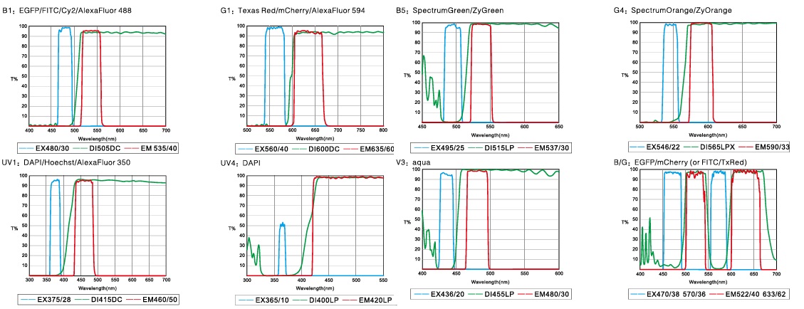
The epi-fluorescence system directs excitation light through the objective, separates excitation and emission wavelengths, and sends the fluorescent signal to the eyepiece or camera.
Fluorophores and applications
Final excitation and emission specifications should always be matched to the actual fluorophores used by the laboratory.
| Fluorophore or method | Typical application |
|---|---|
| DAPI | Nuclear and DNA staining |
| FITC / GFP | Green fluorescence and fluorescent-protein imaging |
| TRITC / Cy3 | Red or orange fluorescence |
| Cy5 | Far-red fluorescence |
| Alexa Fluor dyes | Multicolour fluorescence |
| FISH probes | Chromosome and genetic analysis |
Research applications
Observe labelled cells, proteins, nucleic acids, intracellular components and molecular targets.
Image fluorescently labelled antibodies, cell markers and tissue targets with suitable filter sets.
Support chromosome imaging, fluorescent probes, karyotyping workflows and genetic analysis.
Combine conventional brightfield observation with fluorescence-based specimen examination.
Visualize microorganisms, cellular structures and fluorescent markers.
Configure the optical platform for neuroscience, developmental biology and other research applications.
Technical information
Choose from manual, fluorescence, digital and motorized components to create the right configuration for your application.

PL10X/25 mm high eye-point, wide-field plan eyepiece with diopter adjustment; reticle version available.
PL10X/26.5 mm high eye-point, wide-field plan eyepiece with diopter adjustment; reticle version available.
Plan semi-apochromatic fluorescence objectives: 4X, 10X, 20X, 40X and 100X.
Infinity plan achromatic objectives: 2X, 4X, 10X, 20X, 40X and 100X.
Infinity plan phase-contrast objectives: 10X, 20X, 40X and 100X.
LMI inverted-image 30°/25° inclined Siedentopf trinocular head; 50–76 mm interpupillary distance; R:T ratios of 100:0, 20:80 or 0:100.
Erect-image 30°/25° inclined Siedentopf trinocular head; 50–76 mm interpupillary distance; R:T ratios of 100:0 or 0:100.
187 × 168 mm double-layer mechanical stage; 80 × 55 mm movement range; 0.1 mm precision; two-way linear drive with adjustable tension.
Optional 175 × 145 mm rotatable, anti-corrosive and anti-friction stage; right- or left-hand X/Y controls; 76 × 50 mm movement range.
Optional 187 × 166 mm stage with 80 × 50 mm movement range and 0.1 mm precision.
Biological transmitted-light frame with low-position coaxial coarse and fine adjustment; 25 mm coarse range and 0.001 mm fine precision.
Fluorescence transmitted-light frame with the same focus controls. Both versions include 100–240 V AC, 50/60 Hz wide-voltage transformer, digital intensity setting/reset and LBD, ND6 and ND25 filters.
UV2 long-pass LED module, 365 nm; B4 TB module, 455 nm; B1 band-pass module, 470 nm; G1 band-pass module, 560 nm.
Each module includes intensity control and bright-field/fluorescence switching. Additional modules can be tailored to clinical diagnostic requirements.
100–240 V AC, 50/60 Hz input with 3 W high-brightness LED and continuous intensity adjustment.
Optional 6 V / 30 W halogen bulb with preset filament centre and continuous intensity adjustment.